New Eye Structures Discovered
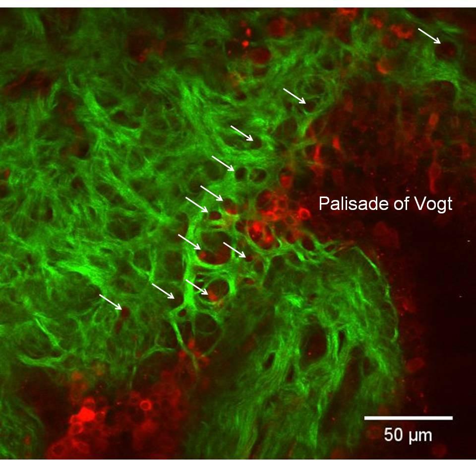
Newly discovered structures within the limbus, the transition zone between the clear cornea and the opaque sclera. Photo: Chuck lab.
"It's not everyday that one newly discovers parts of the human body," says Roy S. Chuck, MD, PhD, Chairman Department of Ophthalmology and Visual Sciences, Albert Einstein College of Medicine.
Yet that's exactly what he and his lab team, in collaboration with Professor Choul Yong Park, a visiting scientist on sabbatical from Dongguk University, South Korea, announced in a recent study published in Investigative Ophthalmology and Visual Science. New structural anatomy is rarely uncovered these days.
"What makes this finding extra interesting is the proximity of these new structures to a stem cell region where we already perform limbal stem cell transplants to replenish the corneal epithelium when it is lost to disease or injury," he adds.
Using an advanced spectroscopic technique (second harmonic generation imaging microscopy), the Chuck lab was able to describe two novel structures in the cornea of the human eye: the anterior limbal cribriform layer and presumed anchoring fibers. These structures have never been described by any means before.
According to Chuck, an Unrestricted Grant from RPB to the Albert Einstein Department of Ophthalmology and Visual Sciences supported user time on the core microscope (NIH and institutional grants covered purchase of the microscope, but major users must cover the upkeep), paid for the purchase of sample preparation materials, and covered manuscript submission and publication costs.
While the function of the collagen/elastin-containing anterior limbal cribriform layer remains unknown, it may play a supportive function as it is composed of structural proteins, passes vasculature and underlies a stem cell region of the eye.
"Hopefully we will be able to better understand the function of these newly discovered structures by monitoring their appearances in various disease states of the ocular surface," says Chuck. "Perhaps they are necessary to maintain the stem cell and/or vascular surrounding environment in healthy corneas."
September 29, 2015
Related News: Cornea , Top Story

New study shows that diabetic eye disease is up, but the most severe forms are down
Data from an RPB and MTMVI-supported study showed that while diabetes-related eye diseases doubled since 2014, the most severe forms of the disease have decreased.

RPB Hosts Vision Research Funding Partnership 2025
Leaders of organizations that fund vision research convened in Washington, D.C. to increase collaboration and maximize the impact of research funding for sight-threatening diseases.

Research to Prevent Blindness Celebrates 65 Years of Success
Since 1960, RPB has been transforming vision research and eyecare for the benefit of all people

Research to Prevent Blindness and Association of University Professors of Ophthalmology Announce 2025 Recipient of RPB David F. Weeks Award for Outstanding Vision Research
Maria Bartolomeo Grant, MD, is recognized for ground-breaking contributions to the field of vision research.

The Time is Now to Protect the National Eye Institute
The existence of the National Eye Institute, the most important source of funding for vision research in the U.S., is being threatened.

RPB Grantees Contribute to Eye Transplantation Effort
The ARPA-H THEA project takes on an exciting challenge.
Subscribe
Get our email updates filled with the latest news from our researchers about preventing vision loss, treating eye disease and even restoring sight. Unsubscribe at any time. Under our privacy policy, we'll never share your contact information with a third party.
| General Info | Grants | News & Resources |
Research to Prevent Blindness
360 Lexington Avenue, 22nd Floor
New York, NY 10017
(212) 752-4333
Copyright © 2025 Research to Prevent Blindness. All Rights Reserved.